708-429-4887
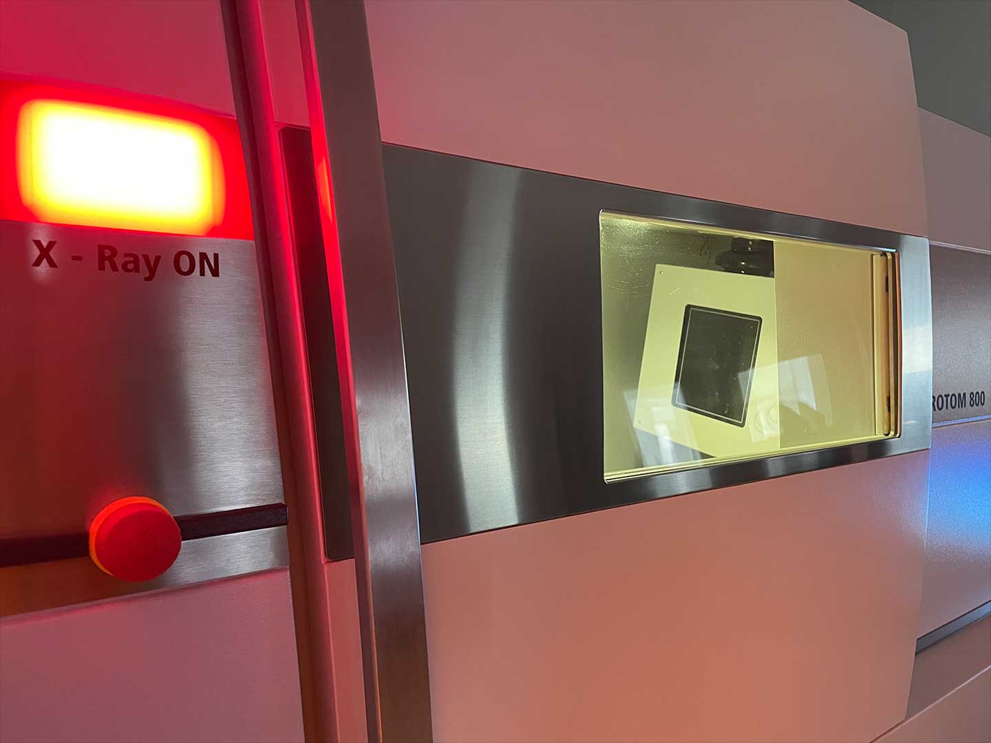
Learn how Nel PreTech achieves accurate and reliable imaging results on thicker, more dense material.
X-ray imaging has revolutionized various fields, from medical diagnostics to industrial inspection. However, as X-rays penetrate thicker materials, they encounter phenomena like scattering and beam hardening, which can compromise image quality and accuracy. In this article, we'll delve into these phenomena, exploring their effects on imaging and discussing strategies to mitigate them through optimization of scan parameters and calibration techniques.
As X-rays traverse through a material, they interact with its atomic structure, leading to scattering and absorption. Scattering occurs when X-rays deviate from their original trajectory due to interactions with the atom’s electrons. This phenomenon causes blurring and distortion in the resulting image, diminishing clarity and fidelity. Beam hardening, on the other hand, arises from the differential absorption of X-rays across a spectrum of energies. As X-rays penetrate a material, lower-energy photons are preferentially absorbed, resulting in a hardened beam with higher average energy. This leads to variations in X-ray intensity across the image, affecting density measurements and material characterization.
.jpg)
.jpg)
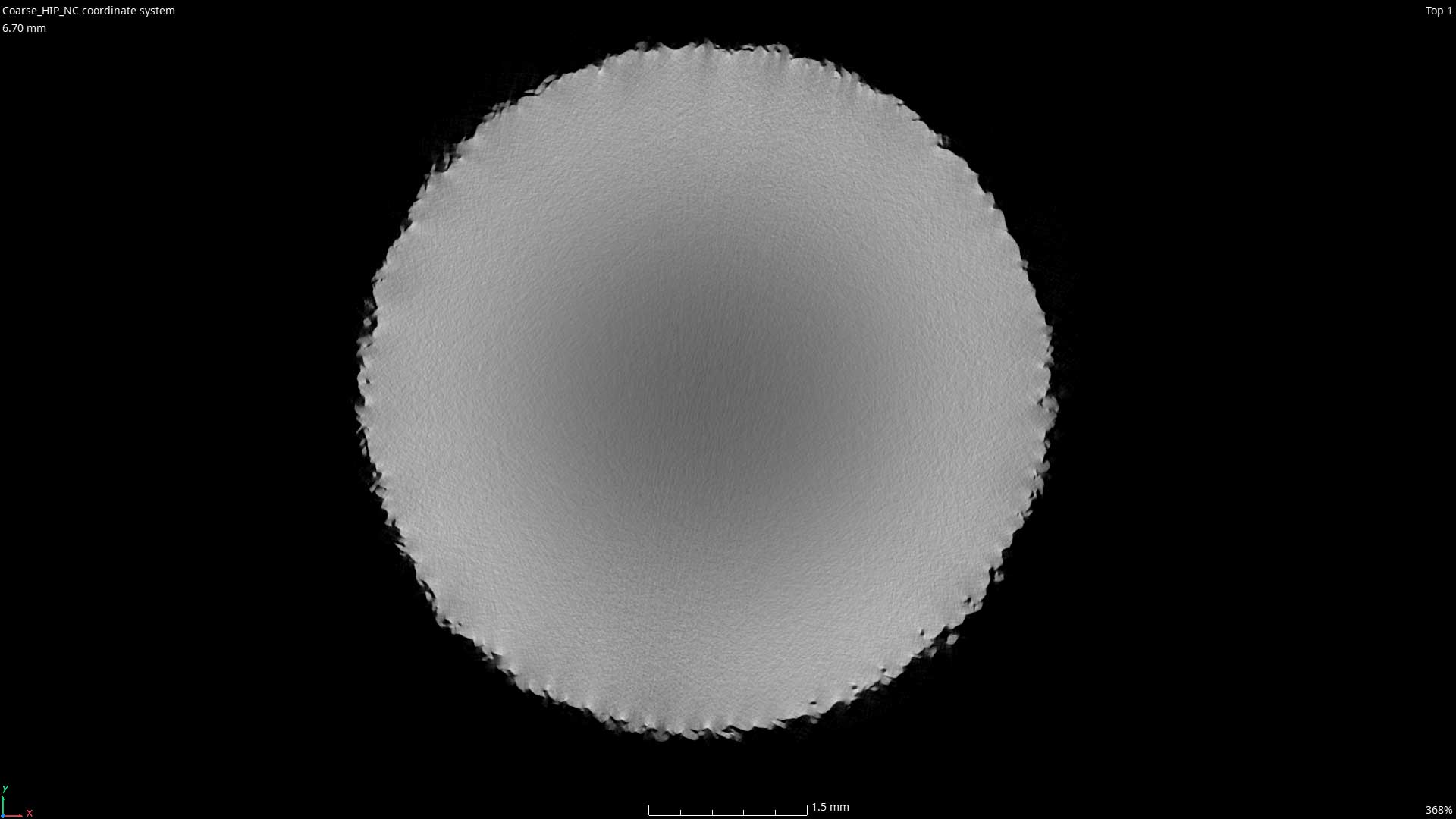
Both scattering and beam hardening effects become more pronounced with increasing material thickness and density. Thicker materials provide more opportunities for X-ray interactions, exacerbating scattering and beam hardening, while higher densities increase the frequencies of the interactions. Consequently, imaging thicker or more dense materials poses greater challenges in terms of maintaining image quality and accuracy. This is particularly critical in fields such as medical imaging and non-destructive testing, where precise characterization of internal structures is paramount.
To mitigate the adverse effects of scattering and beam hardening, optimizing scan parameters is crucial. Key parameters to consider include:
1. Energy Spectrum: Adjusting the energy spectrum of the X-ray source can help minimize beam hardening effects. Using a broader spectrum or employing filters to tailor the energy distribution can yield more uniform X-ray attenuation across the object, reducing artifacts caused by beam hardening.
2. Exposure Time: Optimizing exposure time ensures sufficient signal-to-noise ratio while minimizing radiation dose. Longer exposure times may enhance image quality by capturing more photons, but they can also increase the likelihood of scattering. Balancing exposure time with radiation dose is essential to achieve optimal imaging results.
3. Beam Geometry: The angle and orientation of the X-ray beam relative to the object can influence scattering and beam hardening. Adjusting beam geometry, such as the incident angle and collimation, can help optimize imaging conditions and minimize artifacts.
In addition to optimizing scan parameters, calibration techniques play a crucial role in mitigating scattering and beam hardening effects. Calibration phantoms, consisting of known materials with defined density and composition, are used to calibrate X-ray systems and correct for artifacts. By scanning these phantoms under various conditions, such as different energy levels and thicknesses, engineers can characterize and compensate for scattering and beam hardening artifacts in the imaging system.
Scattering and beam hardening pose significant challenges in X-ray imaging, particularly when imaging thicker and more dense materials. Understanding these phenomena and their impact on image quality is essential for engineers and technicians involved in X-ray-based applications. By optimizing scan parameters and employing calibration techniques, it's possible to mitigate these effects and achieve accurate and reliable imaging results. As technology continues to advance, ongoing research and innovation in X-ray imaging will further enhance our ability to overcome these challenges and unlock new possibilities across diverse domains.
Would you like to speak to one of our experts about x-ray scanning, imaging, or analysis? Contact us today.

You'll find all the detailed service information you need in one brochure.
Download Brochure
Get a quote within 24-hours and keep your project on schedule.
Get a Quote
Our Nel PreTech engineers are ready to get started on your product challenges.
Ask an Engineer